Dry eye disease (DED) is a prevalent and chronic condition, affecting approximately 16 million adults in the United States alone, with meibomian gland dysfunction (MGD) being a leading cause. At the core of MGD management is the practice of manual meibomian gland expression, which is essential for addressing the root cause of this condition. Understanding why this intervention is necessary requires a look at the biology of the meibomian glands, the pathophysiology of MGD, and the scientific rationale behind manual expression.

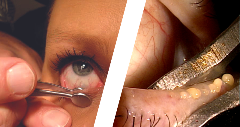
Want to express meibomian glands like a pro? Here’s a step-by-step tutorial.
By Jonathan Hatley, OD, and Nathan Lighthizer, OD
The Role of Meibomian Glands in Ocular Health
The meibomian glands, located in the eyelids, are responsible for secreting meibum, an oily substance that forms the outermost layer of the tear film. This lipid layer is critical for preventing the rapid evaporation of tears, stabilizing the tear film, and maintaining eye comfort. When functioning optimally, meibomian glands release a clear, fluid meibum that spreads evenly across the eye’s surface with each blink.
However, a variety of factors—including aging, systemic health conditions, and environmental influences—can impair meibomian gland function. In MGD, these glands often become blocked, leading to thickening or stagnation of meibum, disrupting normal glandular function, and contributing to dry eye disease. Studies show that up to 86% of patients with dry eye disease have some form of meibomian gland dysfunction, making it a primary cause of tear film instability and discomfort.
The Pathophysiology of MGD
MGD is characterized by the obstruction of the meibomian glands. Over time, glandular ducts may become clogged with keratinized cells, debris, or thickened meibum, restricting the flow of oil into the tear film. This blockage worsens inflammation and disrupts the natural balance of the ocular surface.
Increased viscosity of meibum, often described as resembling toothpaste, is a hallmark of MGD. As meibum becomes more viscous, it becomes increasingly difficult to express. This thickening contributes to the symptoms of dry eye disease. According to one study, over 60% of individuals with MGD report experiencing moderate to severe symptoms, including irritation, redness, and discomfort, which worsen if left untreated. Without intervention, this dysfunction can lead to chronic dryness, irritation, and potential gland atrophy.
Why Manual Expression Matters
Manual meibomian gland expression plays a critical role in addressing the obstructions present in MGD. By applying targeted pressure to the eyelids, trained healthcare professionals can evacuate the thickened meibum, allowing the glands to return to normal function. The scientific rationale behind manual expression lies in its ability to:
- Clear Blockages: Expression physically removes the obstructive material from the glands. Research indicates that expressing blocked glands can improve tear film stability by up to 30% in individuals with MGD, significantly alleviating dry eye symptoms.
- Stimulate Gland Function: Regular expression of the glands can help stimulate meibomian gland activity, encouraging the production of healthier meibum. Studies have shown that glandular expression, when performed consistently, can increase the secretion of meibum by more than 50%, improving lipid layer function and alleviating symptoms of dry eye.
- Reduce Inflammation: Blocked glands contribute to the accumulation of inflammatory mediators and bacteria, further exacerbating dry eye symptoms. By clearing the glands, manual expression helps reduce inflammation. One study found that patients who underwent manual meibomian gland expression experienced a 40% reduction in ocular surface inflammation, helping to further improve eye comfort and health.
The Necessity of Manual Meibomian Gland Expression
Manual meibomian gland expression is a scientifically supported method for managing MGD and alleviating the symptoms of dry eye disease. By restoring proper meibum flow, expression helps to clear gland blockages, stimulate gland function, and reduce inflammation—each of which is essential for maintaining a stable tear film and improving ocular health. In the context of MGD, this simple but effective intervention is vital for preventing further progression of the condition and enhancing the quality of life for patients.
- Baudouin, C., & Labbé, A. (2011). Meibomian gland dysfunction and dry eye disease: A review of the pathology, diagnosis, and management. Journal of the American Optometric Association, 82(8), 451-463.
- Pult, H., & Riede-Pult, B. (2013). Meibomian gland dysfunction: Pathophysiology, diagnosis, and treatment. Optometry and Vision Science, 90(4), 390-401.
- Craig, J. P., Nichols, K. K., Akpek, E. K., et al. (2017). TFOS DEWS II definition and classification report. The Ocular Surface, 15(3), 276-283.
- Lemp, M. A., & Bron, A. J. (2013). The diagnosis and management of dry eye disease: A twenty-five year review. The Ocular Surface, 11(2), 219-249.
- Pult, H., & Riede-Pult, B. (2014). The efficacy of meibomian gland expression: Evidence from clinical studies. Optometry and Vision Science, 91(8), 880-888.